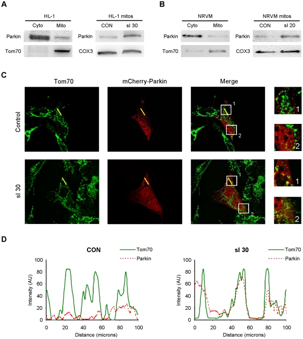
Figure 1. Parkin redistributes to mitochondria in cardiomyocytes subjected to simulated ischemia (sI).
A. HL-1 cells were subjected to simulated ischemia (ischemia-mimetic buffer and hypoxia) for the indicated time, then fractionated to yield cytosol and heavy membranes (mitochondria-enriched fraction). Right-hand panel shows Parkin in the heavy membrane fraction under basal conditions (CON) and after 30 min of simulated ischemia (sI 30). B. Neonatal rat ventricular cardiomyocytes were subjected to simulated ischemia, then fractionated to yield crude cytosol and heavy membrane fractions. Right-hand panel shows Parkin in the heavy membrane fraction under resting conditions (CON) and after 20 min sI. C. HL-1 cells transfected with mCherry-Parkin (red) were subjected to 30 min sI, then fixed and immunolabeled with mitochondrial marker Tom70 (green). Yellow line indicates the segment used for pseudo-line scan analysis. Boxes indicate regions enlarged at right. Images are representative of 5 independent replicates. D. Pseudo-line-scan tracing indicates distribution of mitochondria (Tom70, solid green line) and mCherry-Parkin (dotted red line).

