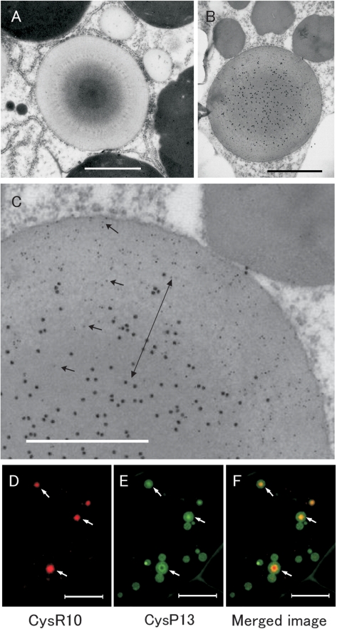
Fig. 1.
Distribution of CysR10 and CysP13 in PB-Is of developing rice endosperm at 3 WAF. (A) Electron micrographs of PB-Is. (B, C) Immunoelectron microscopy of PB-Is which were labelled with anti-CysR10 antibody (15 nm gold particles) and anti-CysP13 antibody (5 nm gold particles). (C) Enlarged image of (B). Arrows in C show the 5 nm gold particles (CysP13). The double arrow in C denotes the middle layer containing CysR10 and CysP13. Bars in (A, B): 1 μm, bars in (C): 0.5 μm. (D–F) Immunofluorescence microscopy of PB-Is from developing rice endosperm at 3 WAF. (D) The distribution of CysR10 as visualized using anti-CysR10 and rhodamine-conjugated secondary antibodies. (E) The localization of CysP13 using anti-CysP13 and FITC-conjugated secondary antibodies. (F) Merged image of D and E. Bars in (D–F): 10 μm.

