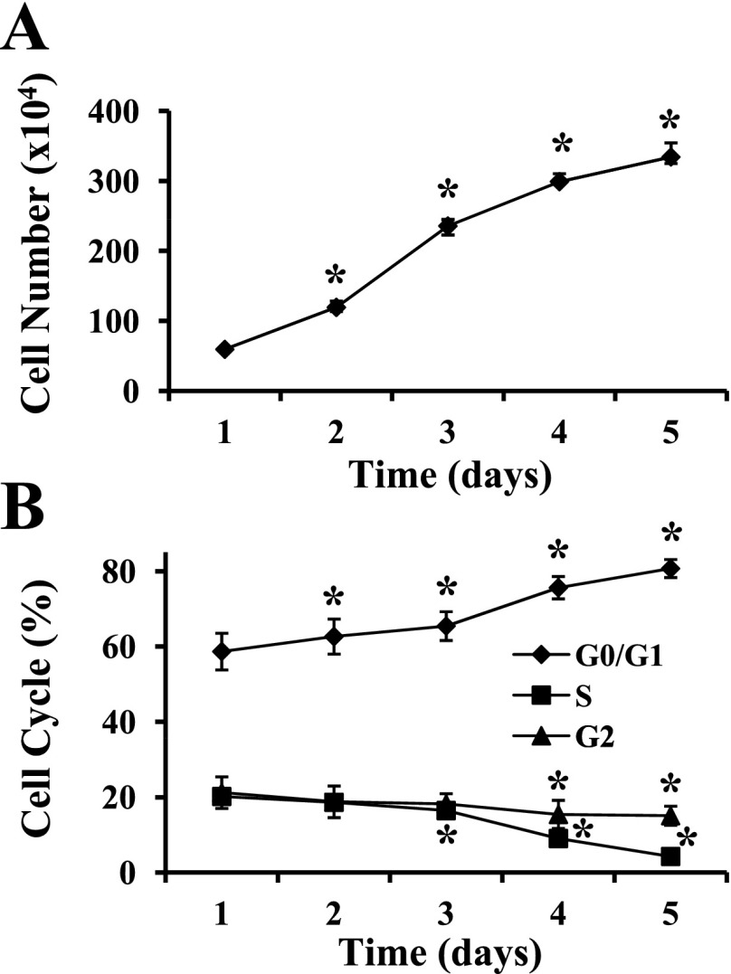
Fig. 1.
Myogenic progenitor cell (MPC) proliferation in vitro. MPC were subcultured after the third passage and maintained in MPC growth medium on type I collagen-coated dishes. A: temporal changes in cell number during proliferation were determined manually. *P < 0.001 compared with proliferation day 1. B: the distribution of cells in different phases of the cell cycle was established by flow cytometry. *P ≤ 0.005 compared with proliferation day 1. Data represent means ± SD of 4 different MPC primary cultures.

