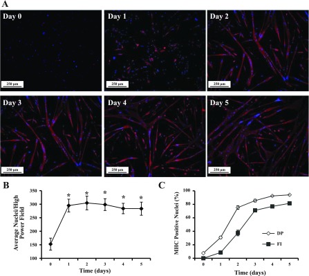
Fig. 3.
MPC differentiation in vitro. MPC were subcultured after the third passage and maintained in MPC differentiation medium on entactin-collagen IV-laminin-coated dishes. A: at various times in culture (day 0 to day 5), differentiated cells were identified by immunolocalization of MHC (red); nuclei were identified by DAPI (blue). B: cell proliferation during the differentiation assay. *P < 0.001 compared with day 0. C: differentiation potential (DP) and fusion index (FI) were determined on the basis of nuclei in MHC-positive cells and myotubes. Data represent means ± SD of 4 different MPC primary cultures.

