
Figure 1.
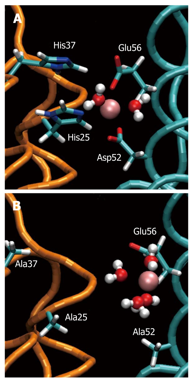
Ferroxidase site of Helicobacter pylori neutrophil activating protein. A: The “ferroxidase site” in the equilibrated wild type. The iron ion (pink) is kept in position by Asp52, Glu56, His25 and His37. Two water molecules are attracted by Fe(II); B: The same site in the equilibrated mutant. The ferrous ion is attracted one-sidedly by Glu56 and Asp53 (not shown) loosing its ability to stabilize the dimer. Four water molecules are attracted by Fe(II).