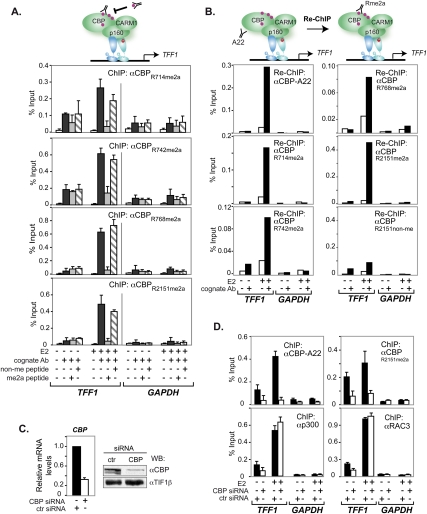
Figure 2.
Methylated CBP was recruited to the TFF1 promoter. (A) H3396 cells, treated with E2 or vehicle (EtOH) for 1 h, were subjected to ChIP with the indicated antibodies. Bound DNA was amplified by real-time PCR with specific primers for TFF1 and GAPDH promoters. For peptide competition, 100-fold molar excess of either the corresponding cognate peptide or an irrelevant control peptide was added to the antibody prior to ChIP. The means ± standard deviations are from at least two independent experiments. (B) H3396 cells were subjected to a first ChIP using the general CBP antibody (A22), and complexes were eluted and subjected to a second immunoprecipitation with or without specific antibody as indicated. Bound DNA was amplified as in A. (C) Knockdown of CBP monitored by RT–PCR (left panel) and Western blotting (right panel). CBP mRNA levels are shown relative to those of 36B4. Immunoblots were done with A22, and TIF1β was the loading control. (D) H3396 cells were nucleofected with siRNAs directed against CBP or a control siRNA (ctr). After 48 h, cells were treated with E2 or vehicle (EtOH) for 1 h and subjected to ChIP with or without the indicated antibodies. Bound DNA was amplified as in A.

