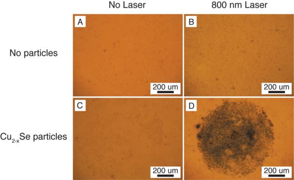
Figure 8.
Comparison of photothermal destruction of human colorectal cancer cells (HCT-116) without (top row – A and B) and with (bottom row – C and D) the addition of 2.8 × 10¹⁵ Cu2−xSe nanocrystals/L. Cells irradiated at 30 W/cm2 with an 800 nm diode laser for 5 min (circular spot size of 1 mm) were stained with Trypan blue to visualize cell death and imaged with an inverted microscope in bright field mode. Significant cell death is observed with 30 W/cm2 irradiation.

