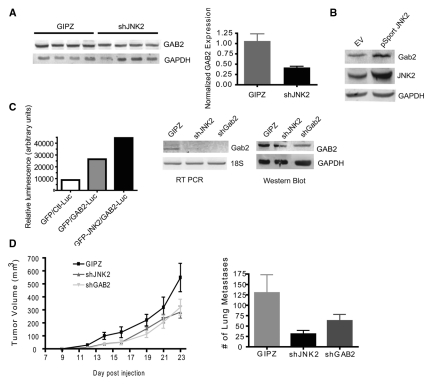
Figure 4.
shGAB2 expression inhibits tumor growth and metastasis. (A) Tumor lysates were evaluated for GAB2 expression using Western blot analysis and normalized to the respective GAPDH levels. (B) Parental 4T1.2 cells were transfected with human JNK2-containing plasmid or empty vector. Cell lysates were then evaluated for JNK2 and GAB2 protein expression using GAPDH as a loading control. (C) Jnk2–/– mammary tumor cells expressing GFP or GFP-JNK2 were transfected with a control luciferase (Ctl-Luc) or a Gab2 promoter luciferase (GAB2-Luc) plasmid along with β-galactosidase. Luciferase absorbances were normalized to β-gal absorbances. 4T1.2 cells were transduced with shGAB2 lentivirus and exposed to puromycin. GAB2 expression was compared among GIPZ, shJNK2, and shGAB2 cell populations using Western blot analysis. GAB2 mRNA expression levels were also measured using RT PCR. (D) GIPZ, shJNK2, or shGAB2 expressing cells were injected into the mammary gland of wild-type mice. Tumors were palpated 3 times weekly until reaching target tumor volume, at which time mice were euthanized (post hoc Student t test, shGAB2 v. GIPZ, P = 0.0167). Lungs were then perfused with India ink, and lung tumor lesions were assessed as described above.

