
Figure 1.
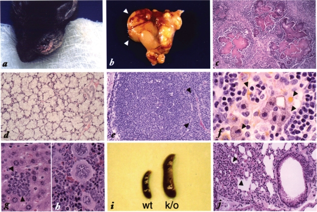
Spontaneous botryomycosis in Pml −/− mice. Pathological features of spontaneous botryomicosis affecting Pml −/− mutants. (A) Botryomycosis in the salivary gland. The left glandular lobe is almost completely replaced by the lesion; overt hyperplasia of the salivary gland is visible (B, D) and also involves the right lobe as indicated by the arrowheads (B). Magnification of the granulomatous area is shown (C): pink cauliflower-like structure consisting of bacterial and proteinaceous material is surrounded by granulocytes and macrophages but mainly by plasma cells. Plasma cell infiltration also affected organs not directly involved in the infection such as the lung and kidney (not shown). (E) Plasma cell proliferation, as indicated by the arrowheads, massively involves the surrounding lymph nodes. (F) Lipochrome granulation of macrophages surrounding the lesion (arrowheads). These cells were also infiltrating the lymph nodes and the lungs (not shown). (G) Extramedullary hemopoiesis in the liver. The arrowheads indicate islands of hemopoiesis. (H) Extramedullary hemopoiesis in the spleen of infected Pml −/− mice (many megakaryocytes are visible), accompanied by massive splenomegaly (I). (J) Interstitial infiltrates of mononuclear cells in the lung. The arrowheads indicate a thickened alveolar wall. Atelectasis of the lung parenchyma is also visible. This picture is frequently observed in apparently uninfected mice.