
Figure 2.
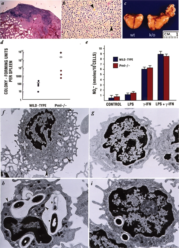
Aberrant response to infections and impaired macrophage function in Pml −/− mice. (A) Massive abscess and atelectasis due to hyperproliferative infiltration of the lung parenchyma in Pml −/− mice infected intraperitoneally with Staphylococcus aureus. Overt peritonitis in 4 of 8 Pml −/− mice was also observed. In control mice, abscesses were confined to the kidney and heart (not shown). In all infected Pml −/− mice, plasma cells and lipochrome macrophages (arrowheads) infiltrated the spleen and lymph nodes. In 3 cases, these cells completely replaced the red pulp of the spleen as shown (B). (C) Hyperplasia of the salivary gland in Pml −/− mice injected locally with Staphylococcus aureus. At postmortem analysis, the injected lobe of the salivary gland of all the Pml −/− was overtly swollen. The injected salivary gland of a representative Pml −/− mutant is shown on the right (k/o). Note the hyperplasia of the left lobe of the salivary gland itself and the hyperplasia of the surrounding lymph nodes that are fused to the lobe. On the left is an injected salivary gland from a control mouse. In this case, the only noticeable alteration is the fibrosis of the injected lobe. (D) Pml −/− mice show impaired response to Listeria monocytogenes infections. The number of Listeria colonies from the spleen of the first of 3 consecutive experiments performed with 4 Pml −/− and 4 wild-type animals is shown. The value obtained from one Pml −/− mutant, which in this experiment spontaneously died at day 5, is indicated by the crossed circle. The 2 mean values from the 2 groups are presented by the dashed open circles. The values obtained from the liver in 2 groups were comparable to the value obtained for the spleen (not shown). Two additional experiments on 8 Pml −/− and 8 wild-type mice, repeated using the same experimental conditions, showed virtually identical results (not shown). The mean values in the 3 experiments showed at least a 100-fold difference between the 2 groups. (E) Nitric oxide production by the Pml −/− macrophages. Resident untreated macrophages were collected from both wild-type and Pml −/− mice and cultured with or without 1,000 U/mL IFNγ, 100 ng/mL LPS, or both IFNγ and LPS for 48 hours. NO2 concentration in the medium was measured with Griess reagent. The experiment shown represents triplicate measurements from the macrophage of 2 Pml −/− and 2 wild-type mice. Three independent experiments showed similar results. (F-I) Defective activation in Pml −/− macrophages is shown (F). Wild-type macrophage upon exposure to Listeria. The plasma membrane is convoluted with pronounced ruffling. The cytoplasm is large and contains numerous lysosomes as indicated by the arrowheads. (G) Pml −/− macrophages upon exposure to Listeria. The plasma membrane is smoother, and the cytoplasm is smaller with few or no lysosomes. (H) Phagocytosing wild-type macrophage. Phagolysosomes with phagocytosed Listeria and fusing lysosomes are clearly visible. The arrowheads indicate lysosomes ready to fuse to the endosome. The asterisk indicates a phagocytosed bacterium, and the open circle indicates a bacterium already partially digested. (I) Phagocytosing Pml −/− macrophage. Phagocytosed bacteria are in vacuoles (see asterisks), but no lysosomes are ready to fuse to them, nor does the macrophage seem to be activated. Magnifications are as follows: 4.400x (F, G) and 10.400x (H, I).