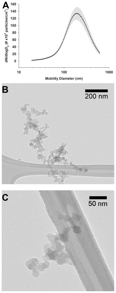
Figure 1.
Particle characterization. Mobility size distribution of the particles in the exposure chamber indicates a geometric mean particle size of 192 nm (A) Values are expressed as mean ± SD. Electron micrographs (B, C) of the particle morphology indicate that the particles varied in shape and that they consisted of primary particles 20–40 nm in diameter and formed larger fractal aggregates.

