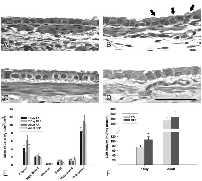
Figure 2.
Morphologic changes in 7 day postnatal and adult rats 24 hours following DFP exposure. Resin sections were analyzed at 60× magnification for histologic changes. (A) In 7 day old postnatal rats reared in filtered air, a simple cuboidal epithelium of mostly ciliated and nonciliated cells was observed in the proximal airways. (B) After DFP exposure, more vacoulated cells were present (indicated by arrows). The epithelium of the large conducting airways was morphologically similar for filtered air (C) and DFP exposed (D) adult rats. Scale bar is 50μm. (E) Cell morphologies of five types: basal, mucous, nonciliated, ciliated, and vacuolated were quantified using point and intercept counts in large intrapulmonray airways. Calculated cell masses are shown (Vs). Although vacoulated cell mass increased and ciliated cell mass decreased following DFP exposure compared to filtered air controls, interactions between exposures were insignificant in both ages. Basal, mucous, and nonciliated cell mass in addition to epithelial thickness remained relatively consistent between exposure groups. Data are mean ± SEM (n=6 rats/group). (F) Lactate dehydrogenase (LDH), a marker for cytotoxicity, was quantified in bronchioalveolar lavage fluid. Significantly more LDH was detected in DFP exposed 7 day old rats, but was not changed in adult animals. Data are plotted as means ± SEM (n=6 rats/group). * = P <0.05, as compared to filtered air controls of the same age.

