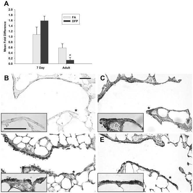
Figure 6.
Gene expression (A) and immunohistochemical staining (B–E) of catalase in the airways of filtered air (B and D) or DFP exposed (C and E) neonatal (B and C) or adult rats (D and E). Gene expression of catalase (A) in microdissected airways was calculated using the comparative Ct method and displayed as a mean fold difference ± SEM (n=3–5 rats/group) compared against FA 7 day postnatal animals using HPRT as the reference gene. After DFP exposure, catalase gene expression remained unchanged in neonates, while a significant decrease in gene expression was found in adults. * = P <0.05, as compared to filtered air controls of the same age. Immunohistochemical staining for catalase protein in FA 7 day neonates was localized to the airway epithelium (B). After DFP exposure (C), catalase staining remained intense within the airway epithelium but increased in other lung compartments. Compared to FA neonates, catalase protein in adult FA exposed rats (D) was increased in abundance. After DFP exposure (E), intense catalase positive staining could be observed within the terminal bronchiole, as denoted by asterisks, in the airway epithelium. Areas with asterisks are pictured in the high magnification insets in D and E respectively. Scale bars are 50μm.

