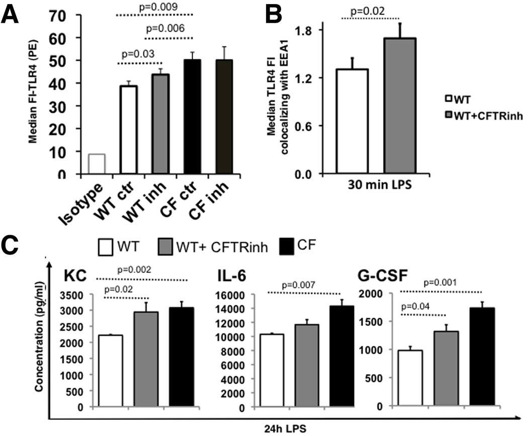
Fig. 6. The abnormal trafficking and signaling in CFTR−/− macrophages is CFTR-dependent.
(A) TLR4 plasma membrane expression as indicated for WT, WT treated with CFTRinh172, CF and CF treated with CFTRinh172 cells: average results for 3 independent experiments (isotype control at left); (B) KC, IL-6 and G-CSF concentration (pg/ml) in the supernatant of LPS treated WT (white bars), WT treated with CFTRinh172 (gray bars) and CF (black bars) macrophages; (C) Average ± SEM MFI of TLR4 localized to the endosomal compartment after 30 minutes of LPS stimulation as assessed by Imagestream for three independent experiments (WT is white, WT treated with CFTRinh172 is light gray). Statistical analysis was performed by two-sample t-tests.

