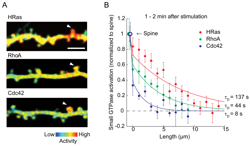
Figure 1. Spatial spreading of small GTPase proteins during structural plasticity.
A. The spatial profile of activity of small GTPase proteins HRas, RhoA and Cdc42 imaged with 2-photon fluorescence lifetime imaging microscopy. Bar: 5 µm. Reprinted and modified from [9] and [11].
B. Activation of small GTPase protein normalized to the stimulated spine as a function of the contour distance from the stimulated spines (Dendritic segment at the spine neck = 0).

