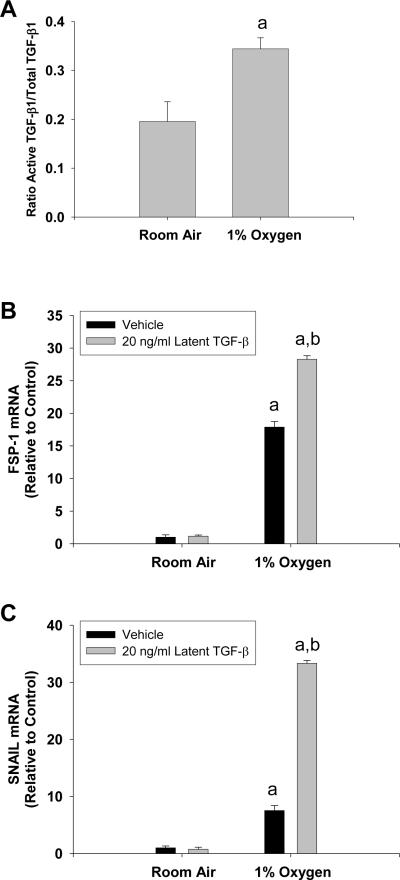
Fig. 10.
Hepatocytes were exposed to room air or 1% oxygen for 24 hours. Active and total TGF-β1 were quantified by ELISA. aSignificantly different from hepatocytes exposed to room air (p<0.05). Data are expressed as means ± SEM; n = 3. Hepatocytes were treated with vehicle or 20 ng/ml latent TGF-β1 followed by exposure to room air or 1% oxygen. 72 hours later, FSP-1 and Snail mRNA levels were quantified. aSignificantly different from hepatocytes exposed to room air (p<0.05). bSignificantly different from hepatocytes exposed to vehicle and 1% oxygen (p<0.05). Data are expressed as means ± SEM; n = 3.

