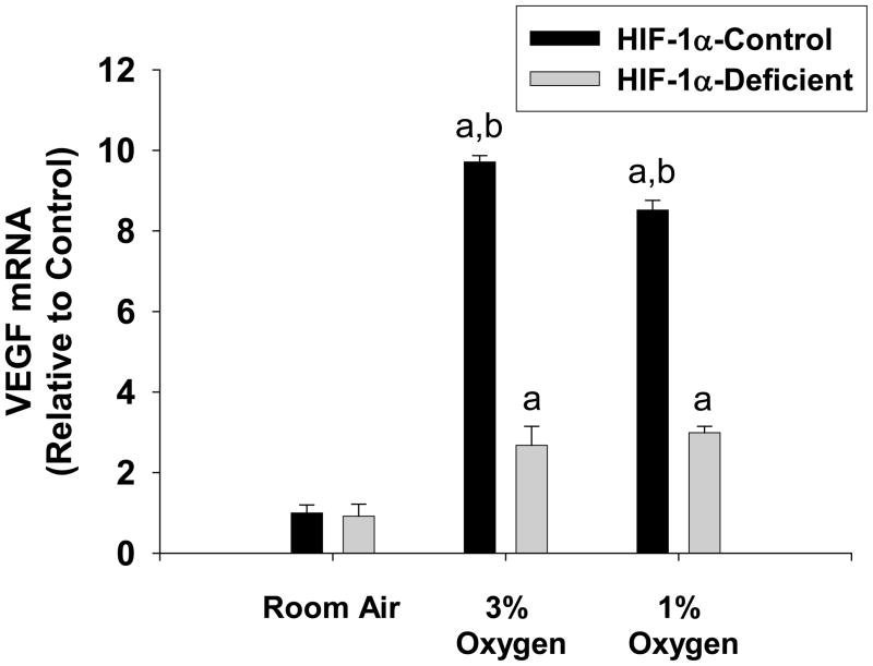
Fig. 5.
Hepatocytes were isolated from HIF-1α-Control and HIF-1α-Deficient mice, and exposed to room air, 3% oxygen, or 1% oxygen. Sixteen hours later, VEGF mRNA levels were quantified by real-time PCR. aSignificantly different from hepatocytes exposed to room air (p<0.05). bSignificantly different from HIF-1α-Deficient hepatocytes exposed to the same concentration of oxygen (p<0.05). Data are expressed as means ± SEM; n = 3.

