Figure 2.
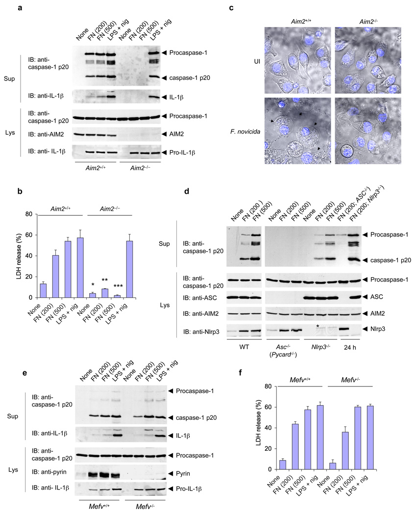
AIM2 is required for F. novicida–induced activation of the inflammasome. (a) Immunoblot analysis of mouse procaspase-1, caspase-1, IL-1β, AIM2 and/or pro-IL-1β in culture supernatants and lysates of mouse Aim2−/− and Aim2+/+ macrophages left untreated or infected for 6 h with F. novicida (FN; MOI in parentheses above lanes) or treated with LPS and nigericin as described in Figure 1b. (b) Release of LDH into culture supernatants of the macrophages in a. *P < 0.05, **P < 0.01 and ***P < 0.005, Aim2+/+ versus Aim2−/− (Student’s t-test). (c) Confocal live-cell microsopy of Aim2−/− and Aim2+/+ BMDMs left uninfected (UI) or infected for 6 h with F. novicida; nuclei were stained with Hoechst stain (blue). Images are merged differential interference contrast and Hoechst channels. Original magnification, x40. (d) Immunoblot analysis of mouse procaspase-1, caspase-1, ASC, AIM2 and/or Nlrp3 in culture supernatants and lysates of mouse wild-type, ASC-deficient (Pycard−/−; called ‘Asc−/−’ here) and Nlrp3−/− macrophages infected with F. novicida for 6 h or for 24 h (far right; MOI in parentheses above lanes). (e) Immunoblot analysis of mouse procaspase-1, caspase-1, IL-1β, pyrin and/or pro-IL-1β in culture supernatants and lysates of mouse pyrin-deficient (Mefv−/−) and pyrin-sufficient (Mefv+/+) macrophages infected for 6 h with F. novicida (MOI in parentheses above lanes) or treated with LPS and nigericin as described in a. (f) Release of LDH into culture supernatants of the macrophages in e. Data are representative of at least three experiments (mean and s.d. in b,f).

