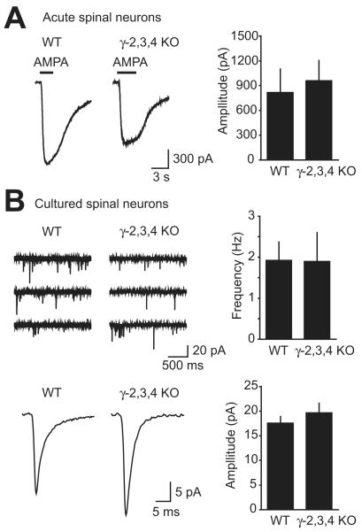
Figure 3. AMPA receptors in spinal neurons from γ-2,3,4 KO mice.
A, Short pulses of AMPA were applied in the presence of cyclothiazide to neurons from E18.5 spinal cord slices. No significant difference in average holding current change was observed between WT, γ-2,3,4 KO, and γ-2,3,4 littermates.
B, Neurons were cultured from WT or γ-2,3,4 KO spinal cords. Typical mEPSC activity is shown by three consecutive 2s sweeps. The average mEPSC for a neuron from each genotype is illustrated. Neurons were included for analysis only if at least 50 mEPSCs were detected. The average amplitude and frequency of mEPSCs was similar in WT and γ-2,3,4 KO mice.

