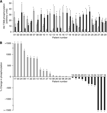
Figure 1.
Phosphorylation of Akt on Thr308 in NSCLC tumour tissue in comparison with patient-matched normal lung tissue. Triplicate samples of lysate from patient-matched normal (N1–3) and tumour (T1–3) tissues were separated on SDS–PAGE gels. Phosphorylation of Akt on Thr308 was determined by western blotting with a pAkt-Thr308 antibody followed by quantitation by densitometric scanning. (A) Quantified data for all 29 patients. Each bar represents the average phosphorylation for normal (N1–3; light grey) or tumour (T1–3; black) tissue for each patient (mean±s.e.m.). The strength of evidence for a difference in phosphorylation between the normal and tumour samples was determined by Kruskal–Wallis test; *P<0.05. (B) The percentage change in Akt-Thr308 phosphorylation in tumour samples in comparison with patient-matched normal tissue where patients are ranked in order of the extent of the percentage change in phosphorylation.

