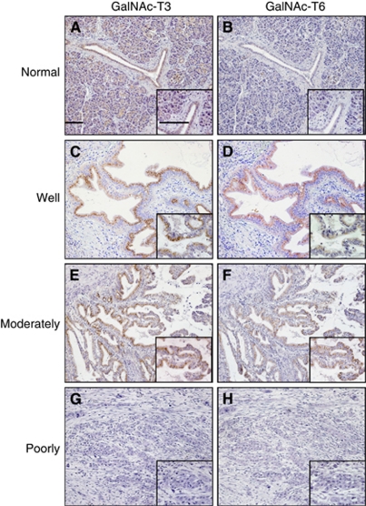
Figure 2.
Immunohistochemical analysis of GalNAc-T3 (1 : 3000 dilution) and GalNAc-T6 (1 : 1000 dilution) in human pancreatic cancers and normal ductal specimens ( × 100; inset, × 400). Bar, 100 μm. Normal pancreatic duct was positive for GalNAc-T3 (A), but negative for GalNAc-T6 (B). Well and moderately differentiated pancreatic cancers stain positively with GalNAc-T3 (C and E) and GalNAc-T6 (D and F), respectively. However, poorly differentiated cancer shows negative staining with both GalNAc-T3 (G) and GalNAc-T6 (H).

