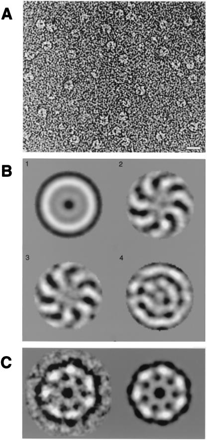
Figure 4.
Sm2⋅U10 complex exhibits, in electron micrographs, a ring structure with a 7-fold symmetry. (A) A typical electron micrograph field of Sm2 in the presence of U10, negatively stained with 2% uranyl formate. (Bar = 1 nm.) (B) Symmetry analysis by multivariate statistical analysis. The first four eigenimages are depicted. The first resembles the total average of the data set. The second and third illustrate both the 7-fold symmetry and the rotational misalignment of the 7-fold symmetric component of the molecular images. The fourth and all subsequent eigenimages (not shown) do not show significant information in addition to the 7-fold symmetry. In particular, residual harmonic components with 6-fold and 8-fold symmetry were not found. (C) Class average of 20 individual molecular images grouped after an automated classification procedure was applied. The class average is shown without (Left) and with (Right) 7-fold symmetry imposed.

