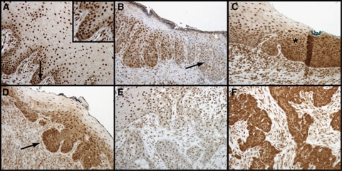
Figure 3.
Characterisation of CIP2A expression during development of oral squamous epithelial dysplasia. (A) Normal oral mucosa with basal cytoplasmic CIP2A positivity (arrow) serving as a control specimen. (B) Severe dysplasia where nuclear positivity disappears (arrow) in oral buccal mucosa. (C) Transition area (asterisk) to carcinoma in situ from the anterior palatinal mucosa. (D) Epithelial budding (arrow) in mucosal tissue transforming into carcinoma. (E) Mild and (F) strong CIP2A immunoexpression in invasive oral squamous cell carcinoma.

