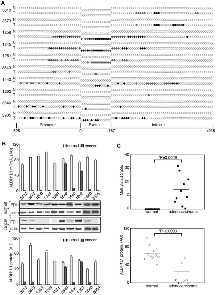
Figure 5.

ALDH1L1 is silenced in human lung adenocarcinomas via methylation within its CpG island. CpG island methylation patterns (A) and alterations in mRNA and protein levels (B) of ALDH1L1 in patients with lung adenocarcinomas. Open circle = nonmethylated CpG; closed circle = methylated CpG; gray circles = CpG methylated in one of the alleles (8 to 14 clones for each fragment were analyzed). Schematic depicts position of CpGs within ALDH1L1 promoter, exon 1, or intron 1. Levels of mRNA were measured by real-time PCR and are presented in arbitrary units (top bar graph). Average ± SD of 3 independent experiments is shown. Protein levels were assessed by Western blot assays; levels of β-actin are shown as loading controls (middle panel). Bottom bar graph: quantification of the Western blots normalized by actin levels. (C) Paired t test analysis of the CpG island methylation (top panel) and ALDH1L1 protein levels (bottom panel) in matching normal and adenocarcinoma tissue samples. Statistically significant differences are indicated by an asterisk. Y-axis in the upper panel shows the number of methylated CpGs (partially methylated sites were assigned the value of 0.5; in all cases, partial methylation was 50% if an even number of clones was analyzed and close to this value if an odd number of clones was analyzed) (Table 3 of Suppl. Material).
