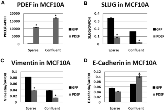
Figure 4.

PDEF expression alters EMT markers in MCF10A cells. Quantitative real-time PCR of (A) PDEF, (B) SLUG, (C) vimentin, and (D) E-cadherin in MCF10A cells plated at low (sparse) and high (confluent) density and transfected with either GFP (black bars) or PDEF (gray bars) normalized to GAPDH. *P < 0.05.
