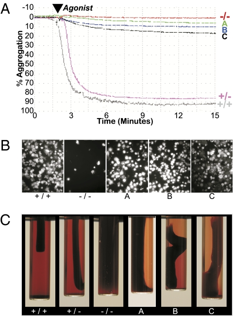
Fig. 2.
Integrin/platelet function was corrected in recipients of αIIb-transduced CD34+ PBC. (A) Aggregation was measured after incubation of washed platelets with fibrinogen and a mixture of activation agonists (ADP, epinephrine, and canine TRAP1, -3, and -4). Platelets isolated from circulating whole blood of αIIb-deficient dogs (−/−) failed to aggregate, whereas the platelets collected from αIIb (+/−, +/+) control animals aggregated to nearly 100% using identical agonist concentrations and reaction conditions. Platelets from αIIb-transduced CD34+ PBC autologous transplant recipients A, B, and C aggregated in direct correlation with the percentage of platelets that expressed αIIbβ3 on their surface (Fig. 1C). Similar results were observed for each transplant dog in at least six separate experiments. (B) An adhesion assay was performed ex vivo to determine whether integrin outside-in signaling initiated by αIIbβ3 interaction with immobilized fibrinogen was corrected in transplant A, B, or C platelets. Washed platelets from αIIb(+/+,−/−) control and transplant dogs A, B, and C were plated onto 100 μg/mL immobilized fibrinogen and allowed to spread for 45 min at 37 °C. αIIb(+/+) platelets bound and spread appreciably onto the fibrinogen, whereas αIIb(−/−) platelets failed to adhere to the fibrinogen. In contrast, transplant dogs A, B, and C platelets bound and spread on fibrinogen-coated plates. This result is representative of three separate experiments. (Magnification, 400×.) (C) Blood was collected from each animal, mixed with human thrombin to allow a fibrin clot to form, and then samples were incubated at 37 °C for up to 24 h. Photographs taken at 3–6 h after addition of thrombin show that normal αIIb(+/+) and carrier αIIb(+/−) samples retracted an appreciable amount of the clot because plasma became readily visible within the test tube. In contrast, the fibrin blood clot remained unchanged within the sample from the αIIb(−/−) GT control dog. In contrast, blood from dogs A, B, and C mediated significant retraction of a fibrin clot similar to the positive controls. This image is representative of the results for dogs A, B, and C from three, seven, and eight separate experiments, respectively, performed for up to 2, 4, and 5 y after PBC transplant, as described in Materials and Methods.

