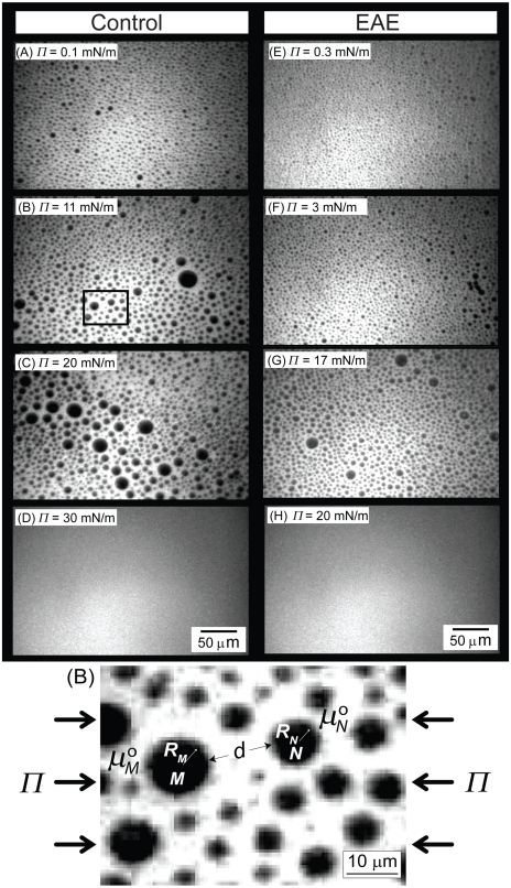
Fig. 1.
Fluorescence images of (A)–(D) control and (E)–(H) EAE CYT myelin monolayers containing 1 wt% TR-DHPE on a MOPS [3-(N-morpholino)propanesulfonic acid] buffer subphase at T ≈ 22 °C and pH ≈ 7.2 as a function of surface pressure. Also shown an enlarged view of domains obtained at 11 mN/m in the control monolayer.

