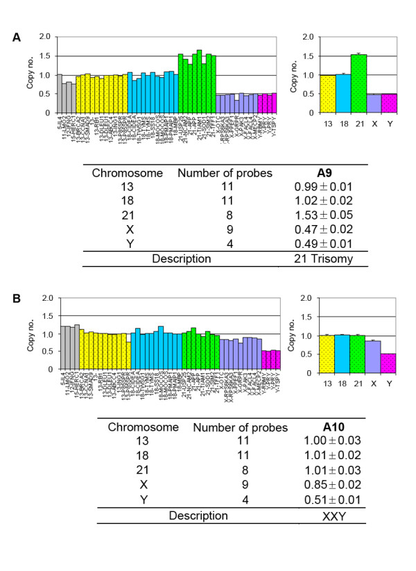
Figure 3.

The male fetus with trisomy 21 and XXY were analyzed with array-MLPA. The left figures showed the relative signal of each probe. The probes on chromosomes 13, 18, 21, X and Y were depicted in yellow, blue, green, purple and purplish red, respectively. Eight autosomal control probes were shown in grey. The right figures showed the average copy numbers on each chromosome. Error bars represented the corresponding SD of the copy numbers on the MLPA probes covering each chromosome. The copy number of each chromosome was listed in the tables.
