Figure 3.
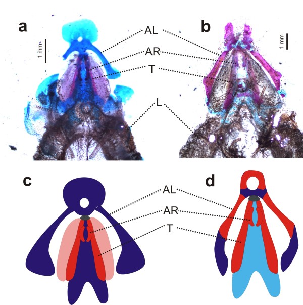
Structure of larynx in Pseudhymenochirus and Hymenochirus. (a) Cleared and stained preparations of the larynx of Hymenochirus boettgeri and (b) of Pseudhymenochirus merlini in dorsal view, showing a generally lower extension of cartilaginous and calcified structures surrounding the larynx in Pseudhymenochirus. L, lungs; AL, alary processes of hyoid plate; AR, arytenoid cartilages; T, thyrohyals (= posteromedial processes of the hyoid plate). Schematic drawings represent main larynx structures in (c) Hymenochirus and (d) Pseudhymenochirus. Colors denote calcified (red) vs non-calcified cartilaginous (blue) structures. Note the calcified alary process in Pseudhymenochirus. Modified from Ridewood [38] and Cannatella and Trueb [29].

