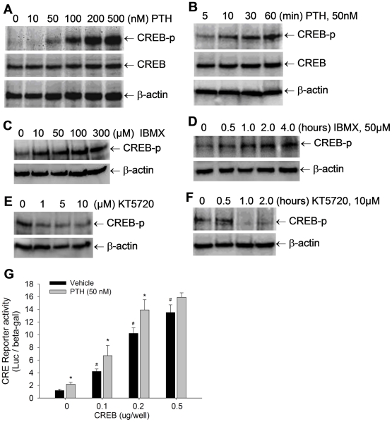
Figure 1. PTH signaling activates CREB phosphorylation in osteoblasts.
(A–F) CREB phosphorylation levels in C2C12 cells, treated with PTH at 0 to 500 nM for 1 hour (A); PTH at 50 nM for 5 to 60 min (B); or IBMX at 0 to 300 µM for 1 hour (C); IBMX at 50 µM for 0 to 4 hours (D); or KT5720 at 0 to 10 µM for 1 hour (E); KT5720 at 10 µM for 0 to 2 hours (F), were detected by Western blot with anti-phosphorylated CREB antibody, with normalization by non-phosphorylated CREB and β-actin. (G) C2C12 cells were co-transfected with CRE-Luc reporter and CREB expression vector, and treated with PTH at 50 nM for 36 hours. The reporter luciferase activity was measured with normalization by β-gal activity. # p<0.01 (CREB vs vector; mean±SE, n = 6); * p<0.05 (PTH vs vehicle; mean±SE, n = 6).

