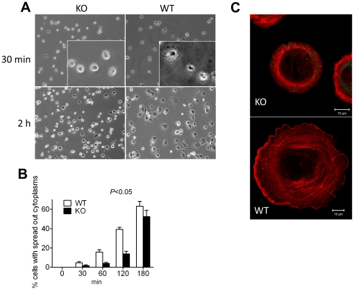
Figure 3. MIM−/− MEFs were impaired in spreading during cell attachment.
(A) Cells were trypsinized, plated on fibronectin-coated coverslips in a serum-containing medium, fixed at 30 min and 2 h and inspected by phase-contrast microscopy. An enlarged area in each 30-min image was shown in inset. (B) The number of cells with apparent cytoplasm extensions was countered at different times after plating. The data represents mean ± SEM (n = 3). The p value (Anova test) refers to the difference between MIM−/− and MIM+/+ cells during the course of attachment. (C) Cells at 2 h after plating were fixed, stained with phalloidin and scanned with confocal microscope with each optical section of 0.30 µm in thickness. The images corresponding to different sections were compiled by Zeiss LSM Image browser.

