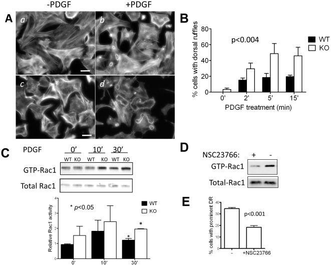
Figure 4. PDGF induces prominent dorsal ruffles in MIM−/− MEFs.
(A) MIM+/+ (a and b) and MIM−/− (c and d) cells were arrested by incubating for 24 h in 0.2% serum-containing medium and then treated with PDGF for 10 min (b and d) followed by staining with phalloidin. The stained cells were inspected by epifluorescent microscopy. Scale bar: 50 µm. (B) Quantification of dorsal ruffles. Cells were treated with PDGF for the times as indicated. The number of cells showing large ruffling areas was counted. The data shown are the mean ± SEM based on four independent experiments. In each experiment 70 cells were analyzed. The p value was calculated by Anova test, referring to the difference between MIM−/− and MIM+/+ MEFs during the response to PDGF. (C) PDGF treated cells were analyzed for Rac1 activation by pull-down assay followed by Western blot using anti-Rac1 antibody. The Rac1 activation was quantified based on three independent experiments. (D) Quiescent MIM−/− cells grown on fibronectin-coated coverslips were treated with and without Rac1 inhibitor NSC23766 at the concentration of 50 µM for 48 h. The treated cells were then stimulated with PDGF for 10 min, and the Rac1 activation was measured as above. (E) NSC23766 treated cells were also treated with PDGF, and the formation of dorsal ruffles (DR) was quantified based on three independent experiments.

