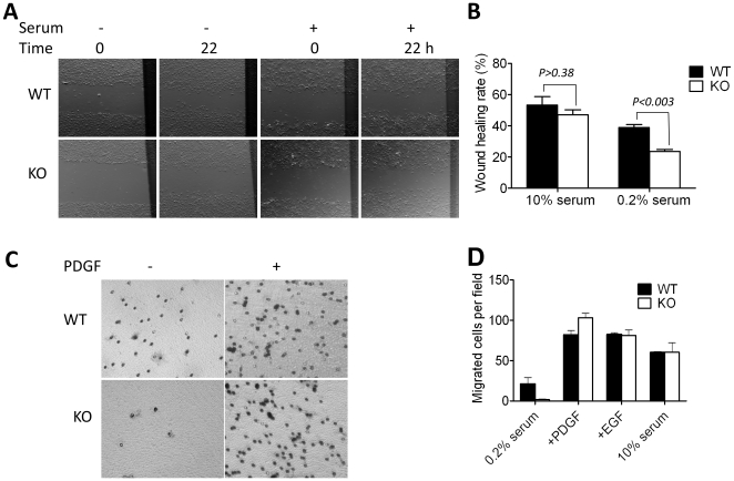
Figure 5. MIM deficiency affects cell motility.
(A) Cells were grown in 10% serum-containing medium until confluence, then wounded by rubber policeman and incubated in medium containing either 10% or 0.2% of serum. The images showing the same wounded areas were taken at the beginning of incubation (0) and 22 h later. (B) Wound-healing rates were measured as described in the Materials and Methods. (C) Cells were arrested in 0.2% serum-containing medium and placed on the top chamber of Transwell plates in which the bottom chamber was filled with 0.2% serum medium or medium containing PDGF (30 ng/ml). The plates were incubated for 4 h, stained and photographed. (D) Quantification of the motility of cells in Transwell plates containing 0.2% serum, 30 ng/ml PDGF, 30 ng/ml EGF and 10% serum, respectively. All the presented values are the mean ± SEM of three independent experiments.

