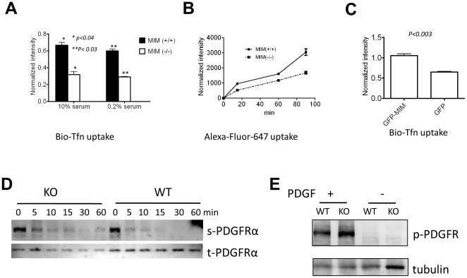
Figure 6. MIM plays a positive role in internalization of extracellular particles.
(A) MEFs were grown in medium supplemented with either 0.2% or 10% serum and incubated with biotin-labeled transferrin (Bio-Tfn) for 5 min. Uptake of Bio-Tfn was measured as described in the Materials and Methods. (B) Cells were treated with Alexa Fluor 647 and incubated for the times as indicated. Internalization of the fluorescent dye was measured by flow cytometry. (C) NIH3T3 cells infected by retroviruses encoding GFP-MIM or GFP only were incubated with Bio-Tfn for 5 min. The internalized Bio-Tfn was measured as described in (A). All the error bars represent the mean ± SEM of three independent experiments. P values (t test) refer to the difference between the samples as indicated. (D) Quiescent MEFs were stimulated with PDGF (50 ng/ml) for the times as indicated. The cell surface (s) and total (t) PDGFRα proteins were analyzed as described in the Materials and Methods. The data represents two-independent experiments. (E) Quiescent MEFs were stimulated with PDGF for 5 min. The total cell lysates were subjected to Western blot using anti-phosphotyrosine antibody (4G10).

