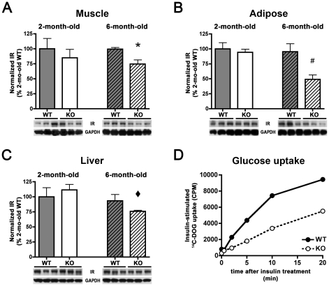
Figure 4. IDE-KO mice show age-dependent reductions in IR levels and insulin-stimulated glucose uptake that correlate with the onset of the diabetic phenotype.
A–C, IR levels in muscle (A) adipose (B) and liver (C) tissue from wild-type (WT) and IDE-KO (KO) mice. Note that IR levels in IDE-KO mice are significantly decreased in all tissues at 6 months, but not at 2 months of age. Graphs show mean ± SEM of IR levels quantified by luminescent imaging (see Materials and Methods ) and normalized to IR levels in 2-mo-old WT mice. *P<0.05 relative to 6-mo-old WT mice; #P<0.01 relative to 2-mo-old KO mice and P<0.05 relative to 6-mo-old WT and KO mice; ⧫P<0.05 relative to 2-mo-old KO mice, all determined by 2-tailed Student's t tests. D, Insulin-stimulated glucose uptake is significantly impaired in primary adipocytes isolated from 6-mo-old IDE-KO mice. Quantification was performed by measuring cellular uptake of 14C-deoxyglucose (14C-DOG) various times after addition of insulin (16.7 nM). Data are mean of 2 independent experiments utilizing epididymal adipose tissue collected from 4 mice per condition for each replication.

