FIGURE 2.
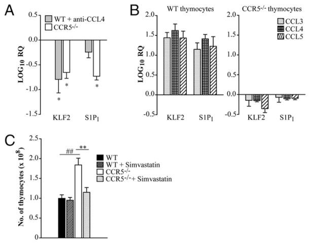
Thymic accumulation is associated with diminished expression of S1P1 and KLF2 in CCR5−/− and CCL4-neutralized mice. Thymi were isolated from WT, CCL4-neutralized, and CCR5−/− mice. Mice were given rat IgG or anti-CCL4 daily for 1 wk days prior to being sacrificed. RNA was extracted from thymi, and expression of KLF2 and S1P1 was measured by quantitative real-time PCR. HPRT was used as an endogenous control, and values represent log decrease compared with WT day 0 thymi (A). WT and CCR5−/− thymocytes were treated with 1 ng/ml CCL3, CCL4, or CCL5 overnight, and KLF2 and S1P1 expression was measured by quantitative real-time PCR. Values represent log change relative to the vehicle control (B). Mice were given 40 μg simvastatin or vehicle control i.p. daily. After 2 wk of treatment, the total number of thymocytes was enumerated (C). Data represent the mean ± SEM (n = 8–12) from two to three experiments. *p < 0.05, **p < 0.01, ##p < 0.001. RQ, relative quantification.

