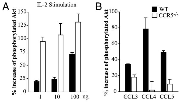
FIGURE 3.
Akt is hyperphosphorylated in CCR5−/− thymocytes. WT and CCR5−/− thymocytes were isolated to measure phosphorylation of Akt. Thymocytes were treated with media alone, 1, 10, or 100 ng/ml IL-2 (A) or 10 ng/ml CCL3, CCL4, or CCL5 (B) for 30 min. Graphs represent the percent increase of total Akt that was phosphorylated after stimulation. Data are representative of two individual experiments.

