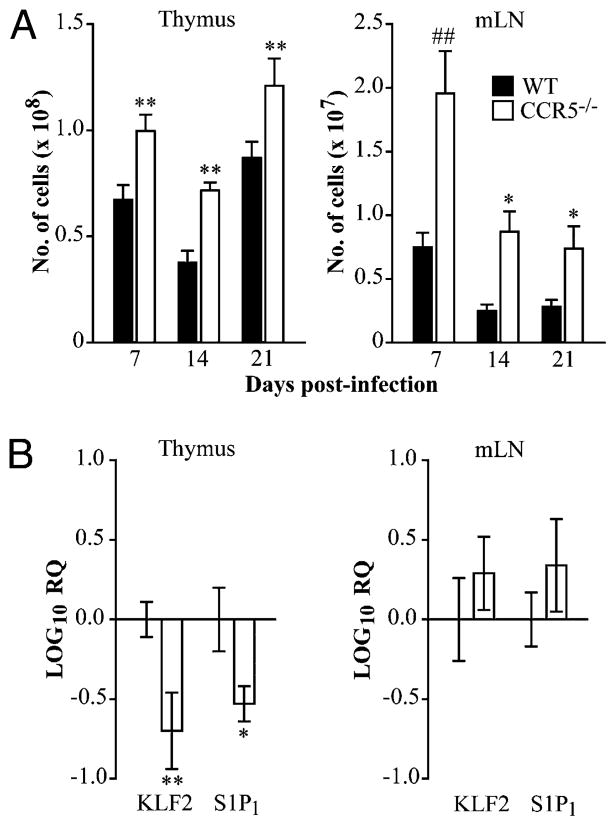
FIGURE 4.
CCR5 influences thymic and lymph node egress during H. capsulatum infection. WT and CCR5−/− mice were infected intranasally with 2 × 10⁶ H. capsulatum and sacrificed at days 7, 14, and 21 post-infection to determine the absolute number of leukocytes in the thymus and mediastinal lymph nodes (A). RNA was extracted from the thymus and mediastinal lymph nodes at day 14 postinfection to measure KLF2 and S1P1 transcription by quantitative real-time PCR. HPRT was used as an endogenous control, and values represent log decrease normalized to WT controls (B). Data represent the mean ± SEM (n = 8–12) from two to three experiments. *p < 0.05, **p < 0.01, ##p < 0.001.

