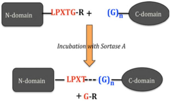
Fig. 1.

Scheme of protein ligation by sortase A enzyme. The consensus sortase residues required at the end of the N-domain are shown (in red) followed by an ‘R’ group (typically His6, in black). Sortase-specific glycine residue(s) required at the N-terminus of the C-domain are in blue. Ligation of both domains results from formation of the new T-G bond (dashed line) that is catalyzed by sortase
