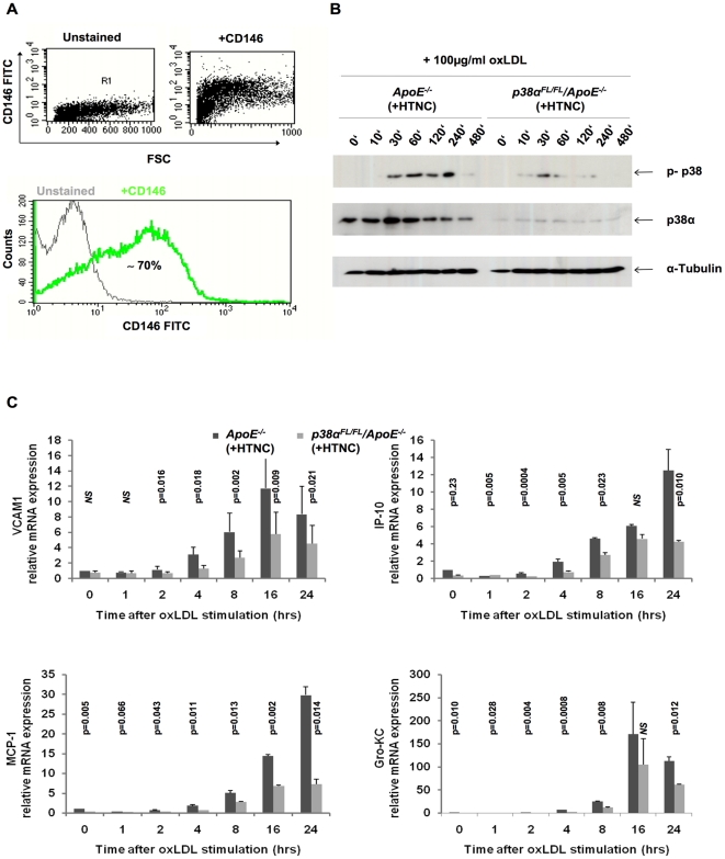
Figure 1. Attenuated monocyte recruitment molecule expression in p38α deficient endothelial cells, in vitro.
(A) Flow cytometric analysis of primary lung endothelial cells to check for purity of population, after treatment with HTNC for 16 hrs. Cells were stained with CD146 FITC, specific for MLECs. Leukocytes isolated from blood were used as negative controls for CD146 staining (data not shown). (B) Immunoblotting for p38α and total p-p38 on MLEC protein extracts. (C) Relative mRNA expression levels of adhesion molecule VCAM1 and chemokines IP-10, MCP-1 and Gro-KC in MLECs. Cells were isolated from the lung of p38αFL/FL/ApoE−/− and ApoE−/− mice, passaged twice and treated with HTNC for 16 hrs to induce cre recombination. Data shown are representative of two separate experiments. Error bars represent SD.

