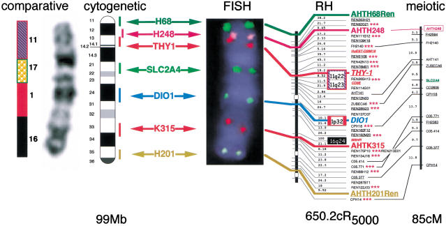
Figure 2.
Multicolor fluorescence in situ hybridization of canine clones to dog chromosome 5 (CFA 5). The integrated RH and meiotic maps for CFA 5 are shown on the right hand side with their corresponding sizes noted below each map. Six clones were selected on the basis of their positions along the length of the RH map and are indicated in colored text. A seventh clone, SLC2A4, was selected on the basis of its position in the meiotic map. Each of the seven clones was labeled with one of the following five fluorochromes: Spectrum Green dUTP (green signal), Spectrum Orange dUTP (gold signal), Spectrum Red dUTP (red signal), DEAC (aqua signal), and Cy5 (pink signal). All seven clones were cohybridized by FISH to elongated dog chromosomes. The resulting FISH image of a DAPI-counterstained CFA 5 is shown in the middle of the figure, illustrating the hybridization signals of all seven clones, along with the assignment of each clone to the DAPI-banded ideogram of CFA 5 (Breen et al 1999a). On the far left is the enhanced DAPI-banded image of the same CFA 5 alongside the corresponding conserved human chromosome segments identified by Breen et al. (1999c).

