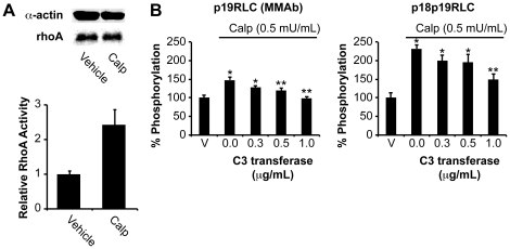
Figure 2. Validation of increased rhoA activity and phosphorylation of RLC by calpeptin.
A. Estimation of GTP-bound ‘active’ rhoA in uterine myocytes treated with Calp or vehicle (DMF) for 15 min (n = 2). RhoA content was quantified relative to α-actin loading control, and used to correct rhoA activity data. Error bars represent standard deviations. B. Quantification of pRLC and ppRLC in uterine myocytes treated with Calp and with increasing concentrations of a cell-permeable rhoA inhibitor (C3 transferase) by ICW (n = 4). * and ** indicate significant differences from vehicle or Calp alone (second histogram), respectively. Calp (calpeptin); rhoA activator (0.5 mU/mL). p<0.05 in all cases as determined by one-way ANOVA followed by Tukey test.

