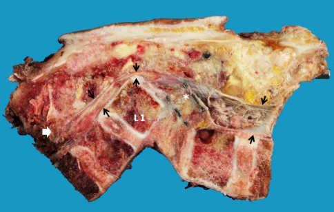
Fig. 7.
The pathological section after en bloc resection, showed the resection margin of the upper and lower vertebral bodies. The dural sac was outlined by the black arrow demonstrated the compression by the postero-inferior corner of L1 body and interbody mesh (asterisk). The epidural invasion of the tumor (white arrow) was completely resected

