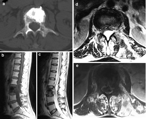
Fig. 3.
Preoperative CT shows cortical destruction of the vertebral body, the left pedicle and the left lamina at L3 axial section (a). Preoperative MRI shows a cystic lesion in the spinal canal at the L2–L3 level with compression to adjacent dura sac, T1-weighted image, sagittal (b), T2-weighted image, sagittal (c), axial section at the L2–L3 level (d). Contrast-enhanced T1-weighted image shows metastatic lesions with contrast enhancement in the left posterior paraspinal region (arrow) at L3 (e)

