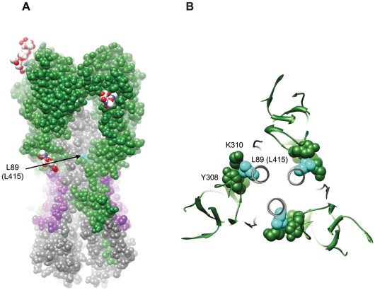
Figure 7. Structural implications for the L89I (L415I) mutation in HA2.
(A) Space filling representation of the surface of A/California/04/2009 HA (PDB:3LZG) to show the recessed location of L89 (L415) (cyan). HA1 is colored green; HA2 is colored gray; the HA2 stem epitope structurally defined in Sui et al. [18] and Ekiert et al. [17] is colored purple; and glycans at glycosylation sites are colored by heteroatom (carbon white, oxygen red, and nitrogen blue). (B) Depiction of the packing interaction between L89 (L415) and residues in an adjacent loop in HA1 (PDB:3LZG). Molecular graphics images were produced using the UCSF Chimera package (supported by NIH P41 RR001081) [45].

