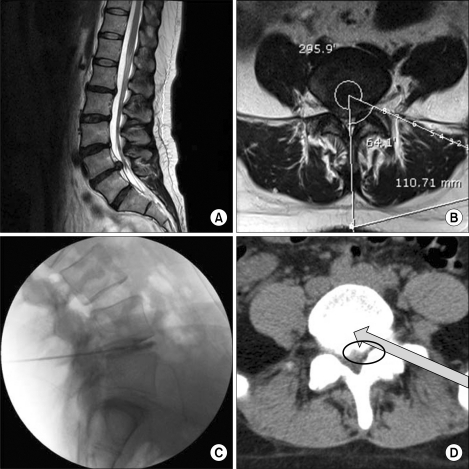
Fig. 1.
The anticipated passage of the needle and working channel while performing percutaneous endoscopic lumbar discectomy at the L4-L5. The anatomic structures from the skin to the targeted anulus at the L4-L5 intervertebral disc space are seen in the following order from the skin surface to the disc: (1) the latissimus dorsi muscle, (2) external and internal oblique muscle, (3) superficial thoracolumbar fascia, (4) erector spinae muscle (lateral tract: iliocostalis lumborum muscle), (5) deep thoracolumbar fascia, (6) quadratus lumborum muscle, (7) erector spinae muscle (lateral tract: intertransversarii mediales muscle), and (8) psoas major muscle. This is a case of a 37-year-old patient who underwent single-level PELD at the L4-L5. (A) Preoperative T2-weighted sagittal magnetic resonance image (MRI); the approaching angle and distance from the midline were measured for the proper placement of the needle and working channel before PELD (B); preoperative T2-weighted axial MRI; (C) intraoperative discogram, lateral view; and (D) postoperative computed tomography. Air shadows are seen in the passage of the working channel in the muscles (arrow) and in the anterior epidural space after the removal of herniated nucleus pulposus using right-angled forceps (circle).

