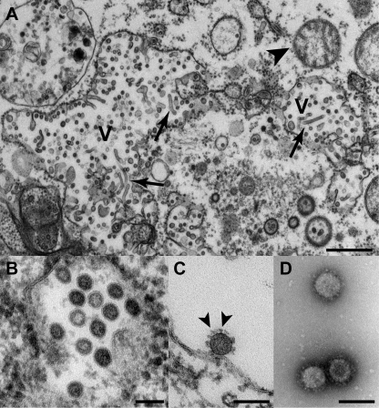
FIG 3 .
CAVV replication and morphology as observed by transmission electron microscopy. Ultrathin sections of C6/36 cells infected with CAVV at 48 hpi (A to C), showing an overview of the cytoplasm of infected cells (A; bar, 1 µm; V, vesicle with virus formation; arrowhead, mitochondrion; arrow, tubular structures likely of viral origin) and a higher magnification of vesicles containing spherical, potentially enveloped particles (B; bar, 100 nm) and separation or adsorption of putative virions on cell membranes (C; bar, 100 nm; arrowhead, spikes on virus surface). Negative staining (1% uranyl acetate) of CAVV sedimented by ultracentrifugation through a 36% sucrose cushion (D; bar, 100 nm). It should be noted that better EM results were obtained at 48 hpi than at 24 hpi.

