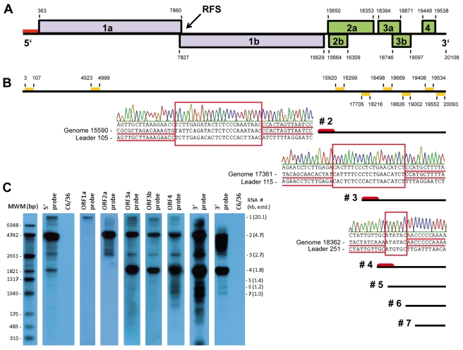
FIG 4 .
CAVV genome organization and subgenomic mRNA synthesis. (A) Positions and sizes of CAVV ORFs. (B) Positions of probes used for Northern blotting and placement of putative sgRNAs. Putative leader sequences are marked by red bars. Electropherograms shown next to mRNAs 2, 3, and 4 indicate typical leader-body fusion sites identified by RT-PCR. No clear leader-body fusions were identified for RNAs shown without leader symbols (see Fig. S2 in the supplemental material). (C) Detection of CAVV genome and sgRNAs by Northern blot analysis of intracellular viral RNA from infected C6/36 cells. Specific probes for the 5′ and 3′ prime ends, as well as for each ORF, were employed. The 3′-terminal probe is shown after short and long exposures of the blot. A molecular size marker (MWM) is shown in the left lane. Molecular size indicators on the right summarize estimated sizes of putative subgenomic mRNAs.

