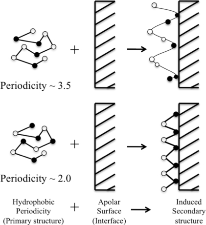
Figure 3.
Schematic diagram showing the effect of hydrophobic periodicity on the secondary structure at interfaces. Filled circles are hydrophobic residues and blank circles are hydrophilic residues. Peptides at apolar/water interface arrange in such a way that will maximize the contact between hydrophobic residues and apolar surface and the contact between hydrophilic surface and aqueous environment (adapted from DeGrado et al. [60]).

