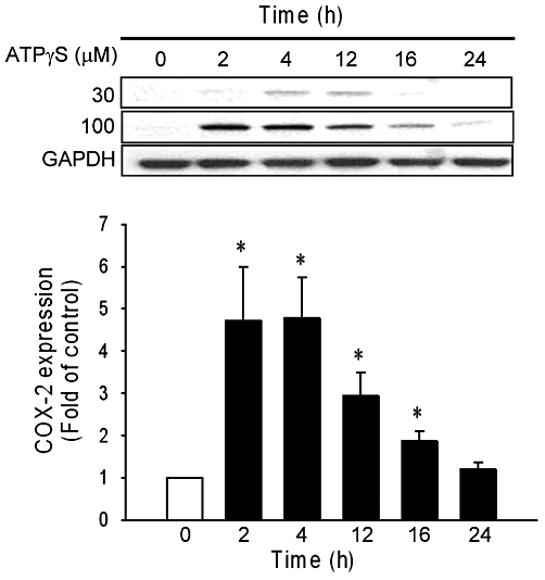
Figure 1.

ATPγS-induced COX-2 expression in vascular smooth muscle cell. Confluent vascular smooth muscle cell were made quiescent for 24 h before incubation with ATPγS (30 and 100 µM) for indicated times. COX-2 expression in response to ATPγS (100 µM) is shown in the lower panel (n = 5). Values are mean ± SE. *P < 0.05 compared with the control group.
